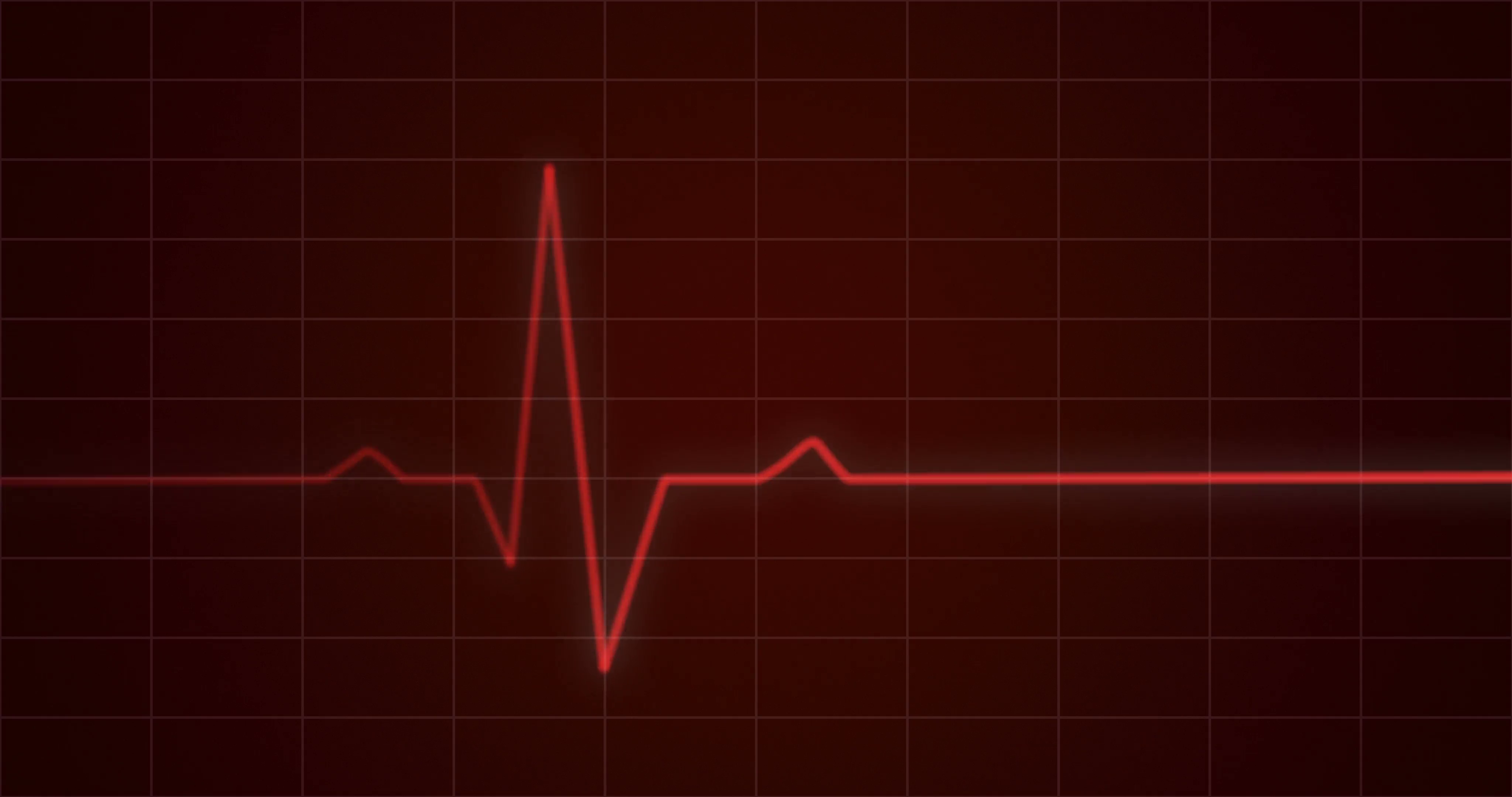
QRS Complex Wave: The “QRS complex,” which is a mix of the Q, R, and S waves, reflects ventricular depolarization. Many cardiac rhythms, including atrial fibrillation and possibly junctional beats, do not have identifiable P waves. Ectopic atrial beats can cause P wave shape to change. Each P wave should be followed by a QRS complex as long as atrial depolarization may propagate through the atrioventricular (AV) node to the ventricles.Ītrial enlargements can broaden or increase the amplitude of the P wave. In this context, the ECG is said to show a normal sinus rhythm. If the action potential is coming from the SA node, the P wave in lead II should be vertical.


When the sinus node, also called the sinoatrial node, produces a nerve impulse that depolarizes the atria, the P wave develops. P Wave: The presence of a P wave suggests atrial depolarization. It is helpful to grasp the theory of what ECGs indicate in order to understand the patterns discovered.Normal beat generates four entities, each with a distinct pattern: P wave, QRS complex, T wave, and a U wave. The reading of the ECG Heart Monitor is essentially a pattern matching process. Few impulses can produce typical variations in cardiac rhythm, while others are potentially dangerous. There can be many hurdles in the transmission of electrical pulse in the system that includes the Atria, SA Node, AV Node or the Ventricles. Some electric pulses do not produce a beat and are responsible for abrupt death. There are also several types of irregular electric pulses, some of which are natural variations and others of which are highly dangerous. The heart typically beats in a sinus rhythm, with each nerve signal produced by the SA node resulting in a contraction of the ventricles, or beat. Tachycardia is defined as a heart rate that is quicker than 100 beats per minute. A heart rate of fewer than 50 beats per minute is referred to as bradycardia. The SA node normally generates an electrical impulse 50-100 times each minute. The doctor can understand well about heart rate and rhythm, and also blood circulation to the ventricles, by analyzing the recording. Several leads provide for a variety of electrical observations of the heart. This electrode leads on the chest by ECG heart monitor can monitor electric pulses produced by the heart. How is ECG Used to Evaluate Heart Function?

Then the right ventricle pumps blood through the pulmonary artery to the lungs and here the carbon dioxide is left off replacing oxygen and so the cycle continues. And so the blood enters the right atrium through the vena cava to get pumped into the right ventricle. When the blood returns, it is depleted of oxygen by carrying carbon dioxide, a waste product of metabolism. Then that blood flows in the left ventricle and is pumped into the aorta to the rest of the body. Through the left atrium, oxygen-rich blood from the lungs enters the pulmonary veins. Our heart has four chambers - right and left atria & right and left ventricles.Blood for our body is collected from the right side and then pumps it to the lungs and the left side of our heart receives blood from the lung to pump it to the body.Blood flows in our body: What Does the Heart Look Like and How Does It Work? There is a standardised ECG heart monitor system that has been developed to position the electrodes for our routine ECG. The electrocardiogram measures the frequency and rate of heartbeats and also provides indirect evidence of blood circulation in the heart muscle. The heart is a dual-stage electric pump and the electrical activity of the core can be measured by electrodes placed on the skin. An ECG heart monitor is a common diagnostic tool used to assess the electrical and muscle functions of the heart.Although this is a simple test to perform, there is a lot of training involved in interpreting the ECG trace.


 0 kommentar(er)
